This project is developing advanced MRI techniques to help us diagnose accurately.

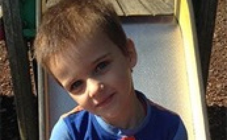
Wilms’ tumour is a cancer of the kidney, mostly affecting children under five. Overall survival rates are high, but there are different sub-types of Wilms’, and we can’t tell which type a child has without surgery. This project is developing advanced MRI techniques to help us diagnose accurately before operating and provide earlier, targeted treatment.

We’re funding an important project to develop advanced MRI scanning techniques, so doctors can treat childhood kidney tumours earlier, with more accuracy, and before surgery.

Thank you

This research project on Wilms’ tumour has been successfully completed. Your donations allow us to fund ground-breaking research that can improve treatments given to children with cancer. Thank you. Your help allows us to continue to find ways to drive up the chances of survival for children with cancer and reduce the toxic side effects that can affect the rest of their lives.

Project Details

  • Project Title

    Advanced magnetic resonance imaging for improved diagnosis and prediction of treatment response in Wilms’ tumour patients

  • Lead Researcher

    Professor Chris Clark

  • Research Centre

    University College London

  • City & Institution Postcode

    London WC1N 1EH

  • Start Date

    7 October 2015

  • Duration

    3 years

  • Grant Amount

    £88,559

Chris Clark

Overview

Wilms’ tumour is a cancer of the kidney. Around 80 children in the UK are diagnosed with it every year – most of them under five. Overall, around 90% survive, but there are different sub-types of Wilms’ and we’re not able to treat all of them so successfully. Children with Wilms’ are given chemotherapy for four to six weeks, to shrink the tumour before surgery. More chemotherapy is usually given after surgery to reduce the risk of the tumour returning. Certain types of Wilms’ tumour respond well to this, but others don’t. Currently we can only find out which type of Wilms’ a child has by examining the tumour after it’s been removed. Then we can see which children have a high-risk type and give them more intensive treatment. If we could identify the tumour sub-type as soon as it’s diagnosed, doctors could tailor treatment sooner, giving the child a better chance of recovery. We’d also know whether the child had a high-risk tumour before we operated, which could reduce the risk of relapse.

What difference will the project make?

Building on a successful pilot study, Professor Chris and his team will explore how an advanced MRI technique known as diffusion-weighted imaging (DWI) could determine a Wilms’ tumour sub-type without surgery, and predict its response to chemotherapy. The team will carry out a DWI scan in each child before and after pre-operative chemotherapy. When the tumour has been removed they’ll examine it to confirm its subtype, and send it for molecular analysis to check for genetic alterations. What they find will show how the tumour has changed as a result of chemotherapy, helping us to understand the differences in how the sub-types of Wilms’ respond. Then the DWI data will be combined with data from the microscopic examinations and the molecular analyses to see if, in future, the imaging data alone could be used to determine which children have a high-risk tumour. In future, this should help doctors decide on the right type of chemotherapy, and plan the timing of surgery based on the child’s individual needs, especially important if both kidneys have been affected. This personalised treatment will help more children with high risk Wilms’ to survive, and also mean that children with standard- and low- risk sub-types can have less aggressive treatment with fewer potential side effects.

About the Research Team

This research is based at the UCL Institute for Child Health (ICH) and Great Ormond Street Hospital (GOSH). GOSH is one of the UK’s major referral centres for children with Wilms’ tumour; the team there sees around a quarter of all Wilms’ patients for surgery. GOSH also receives tissue and imaging scans on all cases registered in the national ‘IMPORT’ study of kidney tumours. Professor Chris Clark is Head of Developmental Imaging and Biophysics Section at ICH. He is a recognised expert in using MRI technology for children; cancer imaging is one of the main themes of his research. His team combines leading experts in paediatric cancer imaging, molecular biology and histopathology, including Professor Kathy Pritchard-Jones and Professor Neil Sebire. Kathy Pritchard-Jones is one of the UK’s leading experts in childhood cancer. She is an Honorary Consultant Oncologist at GOSH and Professor of Paediatric Oncology at ICH, where she heads a laboratory for research into the molecular biology of Wilms’ tumour. Neil Sebire, Professor of Paediatric Pathology at GOSH/ICH, is an internationally established expert in paediatric tumour pathology and is involved in a number of Wilms’ tumour research projects.
Newsletter icon
Newsletter icon

Sign up to our e-newsletter today

Sign up to our e-newsletter and receive exclusive stories straight to your inbox. You will also find out about our latest childhood cancer research news along with updates on our fundraising events, charity news and opportunities to support us. Don’t miss out!

By signing up to this newsletter I agree to receive general and financial appeal emails from Children with Cancer UK