New MRI techniques to improve diagnosis and monitoring of childhood cancer.

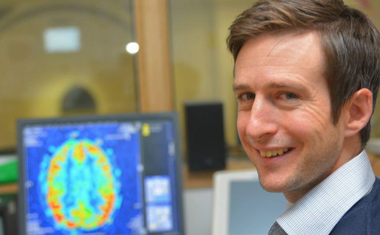
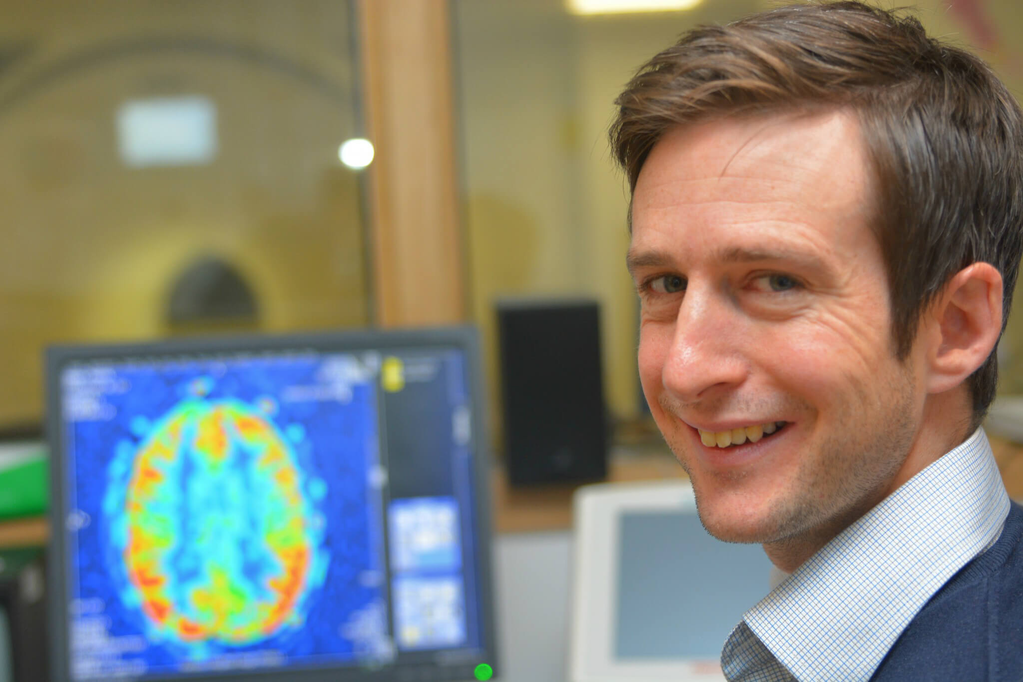
MRI scans are essential to help us diagnose brain cancers. But current techniques can’t always give us the precise information we need about what type or subtype a tumour is, its malignancy, and how far it has spread. Developing new techniques will help us learn more about a brain tumour before we operate.

This Fellowship is funding research into new scanning techniques to help us identify sub-types of brain cancer and give us more information about how to treat them.

Thank you

This research project on new MRI techniques has been successfully completed. Your donations allow us to fund ground-breaking research that can improve treatments given to children with cancer. Thank you. Your help allows us to continue to find ways to drive up the chances of survival for children with cancer and reduce the toxic side effects that can affect the rest of their lives.

Project Details

  • Project Title

    Advanced magnetic resonance imaging techniques for in vivo assessment of tumour heterogeneity, risk factor, and treatment response in paediatric cancer

  • Lead Researcher

    Dr Patrick Hales

  • Research Centre

    Institute of Child Health, University College London

  • City & Institution Postcode

    London WC1N 1EH

  • Start Date

    1 April 2016

  • Duration

    5 years

  • Grant Amount

    £409,310

Patrick Hales

Overview

Some cancers that affect children are still very hard to identify and treat, including the brain tumours high-grade glioma (HGG), diffuse intrinsic pontine glioma (DIPG) and medulloblastoma (MB). As different types of tumour respond better to different treatments, the earlier we can identify them, the better we can plan the right treatment. Magnetic resonance imaging (often called an MRI scan) is essential. But although MRI is good at telling us about where and how big a tumour is, it often can’t tell us enough about it. Also, DIPG and HGG both grow in the brain, but it’s hard to see the details. We need to see how far a tumour has really spread in order to treat it properly. This project aims to develop and test advanced MRI techniques that will help.

Meet the researcher: Patrick Hales

“My hope is that we can use imaging to tell us more and more about actually exactly what type of tumour a patient has and get them onto the right treatment pathway as soon as possible.” Dr. Patrick Hales talks about his research and his motivations in this video.

YouTube needs cookies in order to display videos.
Please Accept Marketing Cookies to watch this video.

What difference will this project make?

To work out the best plan of treatment for a child affected by a brain tumour, we need to know a lot about the cancer, including what type it is. But imaging techniques like MRI can only provide limited information, so we can’t plan individual treatment until after surgery. Yet with certain brain tumours an operation is both difficult and risky. So this project aims to help us find out more at an early stage – before surgery. With new techniques, we’ll be able to identify high-risk tumour subtypes earlier on. We could then see which children may need different treatment and more frequent monitoring. Just as importantly, children with lower-risk tumours might be able to have different therapies with lesser side-effects. Where an operation is needed, knowing more in advance could also guide surgeons to a tumour’s most malignant part. And having more information about where it has spread could help them remove all the tumour tissue without taking out any more healthy brain tissue than necessary, reducing the risk of relapse after surgery as well as the risk of neurological damage. These advanced imaging techniques, which can measure the size of a tumour and give information about its blood supply in a non-invasive way, will also help see how well a child is responding to treatment.

About the Research Team

Dr Patrick will be using a technique called diffusion-weighted MRI (DWI) to get information about the structure of a tumour. A similar technique called diffusion tensor imaging (DTI) will give information about the brain tissue around the tumour. He can also measure the blood supply to a tumour with arterial spin labelling (ASL). He’ll use these with children with brain tumours to see if he can find biomarkers that will help predict what the tumour will do. He and his colleagues will also measure how tumours response to treatment. From ICH, Patrick works closely with clinical colleagues at Great Ormond Street Hospital and Birmingham Children’s Hospital, including Professor Andrew Peet, who is also working with funding awarded under our brain tumour initiative. This means that the imaging techniques can be implemented at two of the largest national referral centres for children with cancer, and taken forward into wider clinical trial.
Newsletter icon
Newsletter icon

Sign up to our e-newsletter today

Sign up to our e-newsletter and receive exclusive stories straight to your inbox. You will also find out about our latest childhood cancer research news along with updates on our fundraising events, charity news and opportunities to support us. Don’t miss out!

By signing up to this newsletter I agree to receive general and financial appeal emails from Children with Cancer UK